Optomap
The most advanced ultra-widefield retinal imagingIntroducing Optomap
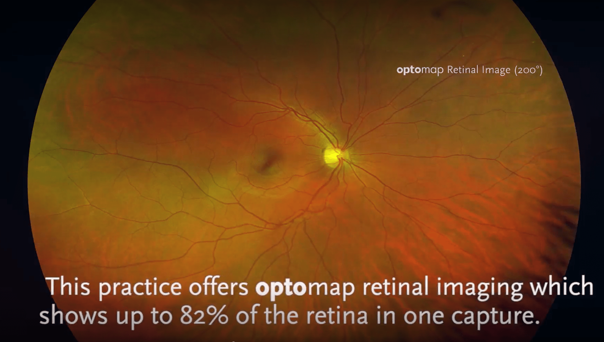
The optomap is a digital image of the retina produced by Optos scanning laser technology. Our Optos device produce ultra-widefield optomap® images of approximately 82% or 200◦ of the retina, something no other device is capable of doing in a single capture.
An optomap image provides a bigger picture and more clinical information which facilitates the early detection, management and effective treatment of disorders and diseases evidenced in the retina.
Now we can see the bigger picture
The fundus is the inside, back surface of the eye. It is made up of the retina, macula, optic disc, fovea and blood vessels. With fundus photography, a special fundus camera points through the pupil to the back of the eye and takes pictures. These pictures help your Optometrist to find, watch and treat disease.
The Optos Daytona uses special technology to capture retinal images as if it were taking them from inside the eye. … The device allows for the discovery of retinal melanomas and peripheral abnormalities such as retinal holes, retinal tears, and retinal detachments. It can also sometimes help us to see vitreal floaters
It means we can now see more of the back of your eye than ever before.
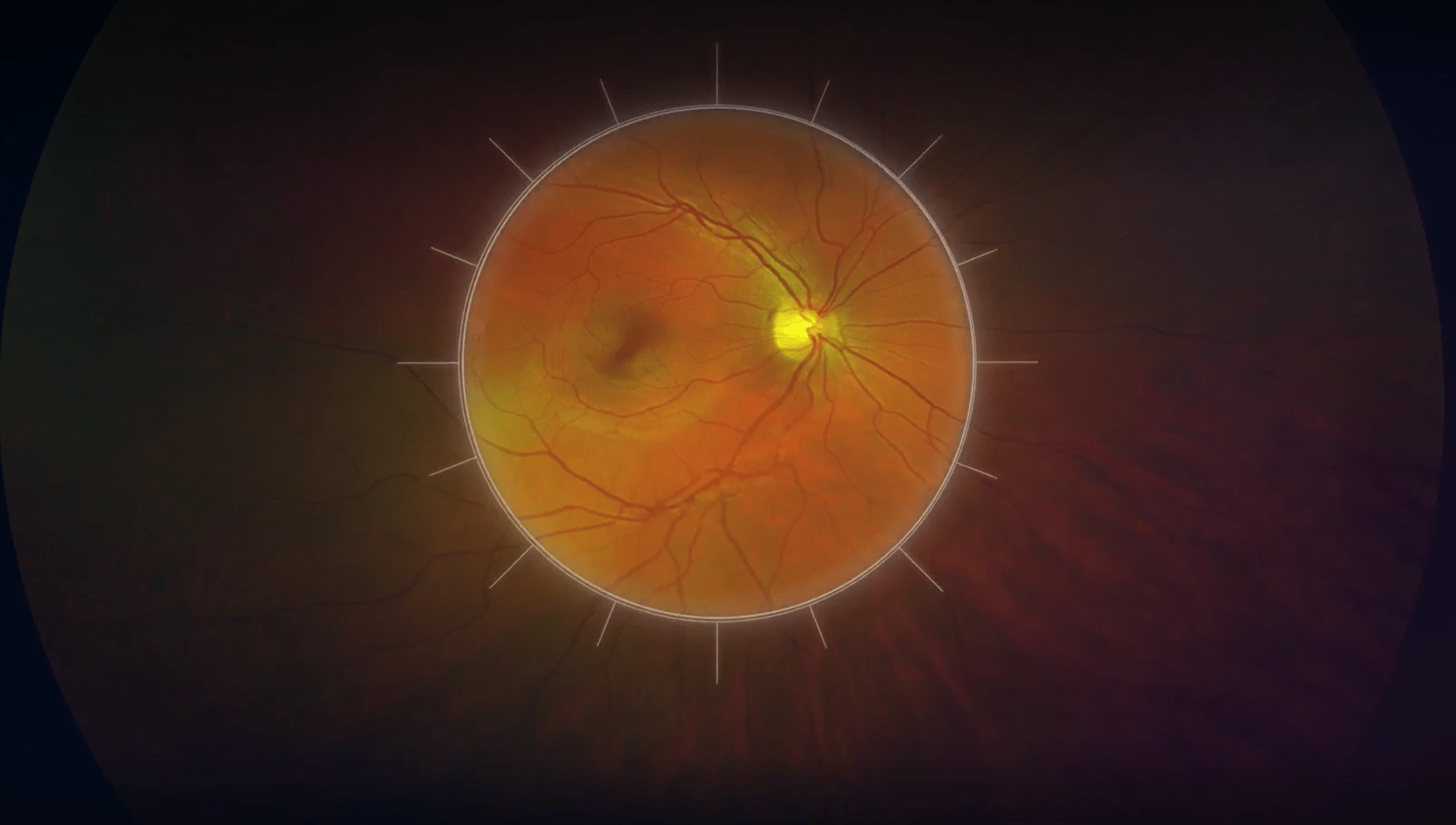
What a traditional retinal photograph shows
A smal part of the back of your eye is visible with a traditional retinal photograph, typically taken on its own before your eye examination with a retinal camera or as an additional part of your OCT scan.
It is useful to the Optometrist in looking at your Optic disk/nerve head which connects your eye to your brain and the Fovea (the focal point) where your important central vision is.
But it doesnt allow the Optometrist to see any more of the back of your eye where other conditions may go undetected. This is where Optomap comes in.
Optomap – a vastly larger area of the back of your eye is shown
The optomap ultra-widefield retinal image is a unique technology that captures more than 80% of your retina in one panoramic image while traditional imaging method typically only show 15% of your retina at one time. Benefits: Early protection from vision impairment or blindness.
The Optomap uses a low-power multi-laser ophthalmoscope to capture digital scans of your retina. It employs the red and the green wavelength to capture retinal images. It filters your image to allow several layers of your retina to be evaluated. And it does it in less than a second! Fast, efficient and accurate.
Optomap
Optomap is the future of digital retinal imaging and it is available at Benjamin Opticians as part of your enhanced eye care examination.
Join Our Conversation


